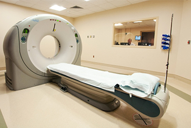
Spiral multidetector CT uses multiple detectors during continuous motion of the patient through the radiation beam to obtain fine detail images in a short exam time. With rapid administration of intravenous contrast during the CT scan, these fine detail images can be reconstructed into three-dimensional (3D) images of carotid, cerebral, coronary or other arteries.
The introduction of computed tomography in the early 1970s revolutionized diagnostic radiology by providing Clinicians with images of real three-dimensional anatomic structures. CT scanning has become the test of choice in diagnosing some urgent and emergent conditions, such as cerebral hemorrhage, pulmonary embolism (clots in the arteries of the lungs), aortic dissection (tearing of the aortic wall), appendicitis, diverticulitis, and obstructing kidney stones. Continuing improvements in CT technology, including faster scanning times and improved resolution, have dramatically increased the accuracy and usefulness of CT scanning, which may partially account for increased use in medical diagnosis.
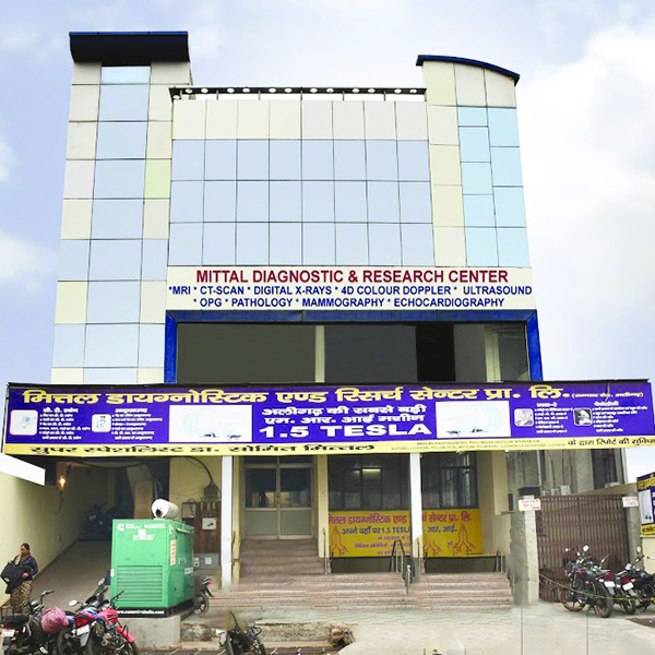
We at MDRC provide whole body imaging in a lesser time frame, with accurate diagnosis.Dual slice spiral CT scanner with 3D reconstruction facility.


