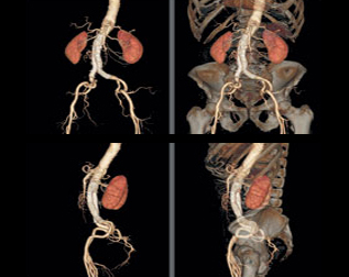
Cross-sectional imaging is usually used to refer to CT, MRI, PET, and SPECT and related imaging techniques, that view the body in cross-section i.e. as axial (cross-sectional) slices.
Ultrasonography is sometimes included under this umbrella term, especially with reference to echocardiography, which produces standardised axial slices through the heart. However most radiologists do not tend to think of ultrasound as a cross-sectional technique since although it generates two-dimensional image “slices” of the body, the angle of the slices are often not perpendicular to the axis of the body.
Examples of imaging techniques that are not cross-sectional include plain radiography, and fluoroscopy. The techniques rely on projection of an x-ray beam through an object to a receptor. Planar nuclear medicine is also not cross-sectional, although the imaging radiation is emitted from the object of interest, rather than passed through it.
Cross sectional imaging really helped us in better understanding of anatomy and various pathologies associated with it.


