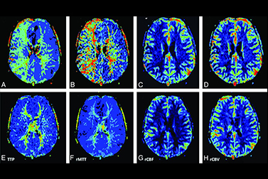
Perfusion weighted imaging is a term used to denote a variety of MRI techniques able to give insights into the perfusion of tissues by blood.
There are three techniques in wide use to derive one or more perfusion values:
Techniques
- Dynamic susceptibility contrast (DSC) MR perfusion.
- Dynamic contrast enhanced (DCE) MR perfusion.
- Arterial spin labelling (ASL) MR perfusion.
Derived values
- Time to peak (TTP)
- Tean transit time (MTT)
- Cerebral blood volume (CBV)
- Cerebral blood flow (CBF)
- Negative enhancement integral (NEI)
- k-trans
The main role of perfusion imaging is in evaluation of ischaemic conditions (e.g. acute cerebral infarction to determine ischaemic penumbra, moya-moya disease to identify vascular reserve), neoplasms (e.g. identify highest grade component of diffuse astrocytomas, help distinguish glioblastomas from cerebral metastases) and neurodegenerative diseases.


