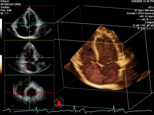
An echocardiogram (echo) is a test that uses high frequency sound waves (ultrasound) to make pictures of your heart. The test is also called echocardiography or diagnostic cardiac ultrasound.
Why do people need an echo test?
Your doctor may use an echo test to look at your heart’s structure and check how well your heart functions.
The test helps your doctor find out:
- The size and shape of your heart, and the size, thickness and movement of your heart’s walls.
- How your heart moves.
- The heart’s pumping strength.
- If the heart valves are working correctly.
- If blood is leaking backwards through your heart valves (regurgitation).?
- If the heart valves are too narrow (stenosis).
- If there is a tumor or infectious growth around your heart valves.
The test also will help your doctor find out if there are:
- Problems with the outer lining of your heart (the pericardium).
- Problems with the large blood vessels that enter and leave the heart.
- Blood clots in the chambers of your heart.
- Abnormal holes between the chambers of the heart.
What are the risks?
- An echo can’t harm you.
- An echo doesn’t hurt and has no side effects.
How do I prepare for the echo?
- You don’t have to do anything special. You can eat and drink before the test like you usually would.
- An ECG (electrocardiogram) records the electrical activity of your heart at rest. … The resting ECG is different from a stress or exercise ECG or cardiac imaging test.
- You may need an ECG test if you have risk factors for heart disease such as high blood pressure, or symptoms such as palpitations or chest pain.


