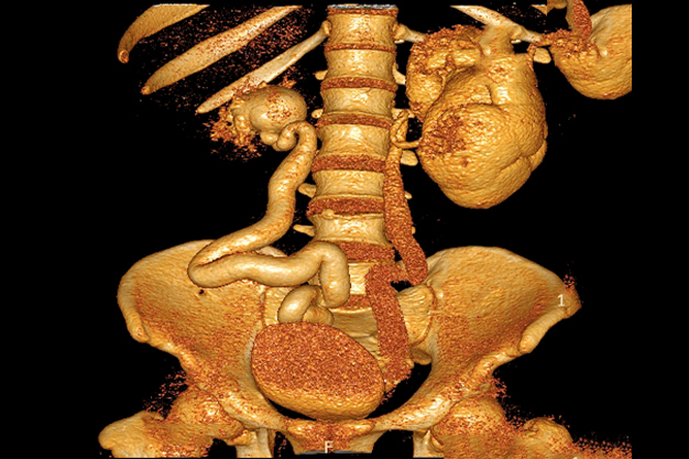
Ectopic insertion of bilateral ureters with proximal hydro-uretero-nephrosis
An ectopic ureter is a congenital renal anomaly that occurs as a result of abnormal caudal migration of the ureteral bud during its insertion to the urinary bladder. Normally the ureter drains via the internal ureteral orifice at the trigone of the urinary bladder.
In females, the most common site for ureter insertion is bladder neck and upper urethra (33%), vaginal vestibule between the urethra and introitus (33%), vagina (25%), and cervix and uterus (<5%).
In males, the ureter may insert into the lower urinary bladder, posterior urethra, seminal vesicle, ductus deferens, ejaculatory duct and rarely the rectum.
Epidemiology
The incidence of ectopic ureters is 1 in 1900, but may be higher because it is often under-diagnosed when asymptomatic, especially in males. It more common in females – F:M = 10:1.
Pathology
Failure of separation of ureteral bud from wolfian duct results in caudal ectopia.
In the case with complete duplication, it obeys the Weigert-Meyer rule – the ureter drains the upper moiety inserts more medial and more inferior to the lower moiety ureter and liable for obstruction while the lower moiety ureter is liable for reflux.
Patients are symptomatic when the ureter inserts beyond the external urethral sphincter.
Associations
may be solitary or a part of complex congenital anomalies, e.g. VACTERL
approximately 80% associated with duplex kidneys
Radiographic features
Intravenous urography (IVU)
It can detect abnormal ureteral insertion and associated anomalies e.g. renal duplication. In complete duplex kidney and ureter, the ectopic ureter usually drains the upper moiety and associated with ureterocele and obstruction.
Voiding cystourethrogram
Usually the ectopic ureter is associated with vesico-ureteric reflux, which can detected and graded with VCUG.
Ultrasound
Associations and complications such as duplex kidneys, hydronephrosis and ureterocoele can be also be assessed.
MR urography
With use of heavy T2 weighted imaging the ureter and its insertion may be visualised. MRI is also useful in detection of other anomalies e.g. renal duplication, ureterocoele and vertebral anomalies
Treatment and prognosis
The more distal the insertion, the worse the prognosis


