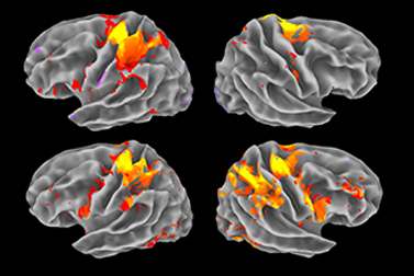
Functional Magnetic Resonance Imaging or functional MRI (fMRI) measures brain activity by detecting changes associated with blood flow. This technique relies on the fact that cerebral blood flow and neuronal activation are coupled. When an area of the brain is in use, blood flow to that region also increases.
The primary form of fMRI uses the blood-oxygen-level dependent (BOLD) contrast. This is a type of specialized brain and body scan used to map neural activity in the brain or spinal cord of humans or other animals by imaging the change in blood flow (hemodynamic response) related to energy use by brain cells
Physicians use fMRI to assess how risky brain surgery or similar invasive treatment is for a patient and to learn how a normal, diseased or injured brain is functioning. They map the brain with fMRI to identify regions linked to critical functions such as speaking, moving, sensing, or planning. This is useful to plan for surgery and radiation therapy of the brain. Clinicians also use fMRI to anatomically map the brain and detect the effects of tumors, stroke, head and brain injury, or diseases such as Alzheimer’s, and developmental disabilities such as Autism etc.


